






Diseases & Conditions
Tenosynovial Giant Cell Tumor (TGCT)
This article was previously titled and focused on pigmented villonodular synovitis (PVNS). Since the World Health Organization now classifies PVNS and giant cell tumor of tendon sheath as tenosynovial giant cell tumors, this article now refers to PVNS as tenosynovial giant cell tumor (TGCT) within joints and has been expanded to include lesions outside of joints (what used to be called giant cell tumor of tendon sheath). These changes were made because tumors within and outside of joints have similar appearance and biology.
Tenosynovial giant cell tumor (TGCT) is a tumor that grows inside the thin layer of tissue that lines the joints (synovium) and tendons (tendon sheaths), causing the tissue to thicken and overgrow.
The tumor is not cancer and does not spread (metastasize) to other areas of the body. However, it is a locally aggressive progressive disease. What does that mean?
- When it involves the lining of joints, TGCT can damage the cartilage and area around the joint, including nearby bone, leading to pain, swelling, and arthritis.
- When it involves the tissue around tendons, TGCT can lead to wearing down of the tendon and surrounding structures.
How Does Tenosynovial Giant Cell Tumor Affect Joints and Tendons?
In a healthy joint, the synovium produces a small amount of fluid that lubricates the cartilage and aids in movement. In TGCT, the synovium is filled with a fragile, vascular tumor that bleeds easily.
In addition to the tumor destroying cartilage and bone, bleeding from the tumor can lead to inflammation in and around the joint, causing swelling and making movement very painful.
TGCT that occurs around tendons can cause them to become thinner and not function properly. In rare instances, tendons can rupture (tear).
TGCT typically involves only one site.
- In 80% of patients who have TGCT within a joint, the knee is involved
- Tumors within joints can also affect the hip, ankle, shoulder, and elbow.
- When TGCT affects tendons, this usually occurs along the wrist, hand, and fingers but can also affect the ankle or foot.
This condition can affect people of all ages, but it occurs most often in young adults from 20 to 50 years of age and is more common in women.
There are two forms of TGCT — localized (also called nodular) and diffuse.
Localized (nodular) TGCT
When the tumor is made up of one or more separate nodules inside of the joint or around a tendon, it is called localized TGCT. This type usually responds well to surgical treatment. The rate of it coming back 5 years after treatment is reported to be 10 to 30%.
Diffuse TGCT
When the condition involves more of the entire joint or spreads along the length of tendons or into the surrounding muscle, it is called diffuse TGCT.
This form of TGCT tends to be more aggressive and is harder to treat. It often requires the input of doctors with backgrounds in different areas (e.g., orthopaedic surgeons, medical oncologists). The reported rate of recurrence 5 years after treatment is 20 to 70%.
Cause of Tenosynovial Giant Cell Tumor
There is no known relationship between environmental exposures or other health conditions and TGCT.
Genetic changes associated with both nodular and diffuse TGCT have been identified. These focus on differences in the colony-stimulating factor-1 (CSF-1) gene. The changes in this gene lead to higher levels of CSF-1, which in turn results in increased numbers of inflammatory cells in the joint or tendon sheath, resulting in swelling and pain.
Identifying this alteration in the CSF-1 gene is not needed to make the diagnosis of TGCT.
Symptoms of Tenosynovial Giant Cell Tumor
Localized TGCT:
- The main symptoms are pain and swelling in the affected joint, due in part to chronic inflammation that the tumor can cause.
- Other symptoms may include stiffness, locking, catching, and instability in the joint, often due to the tumor interfering with the motion of the bones within the joint.
Diffuse TGCT:
- There is often a gradual onset of symptoms including joint pain, swelling, and stiffness.
- Sometimes, patients may experience hemarthrosis, a condition in which blood collects in the joint space. This can occur with little or even no trauma to the affected joint.
In both localized and diffuse TGCT, you may be able to feel a mass, and symptoms may come and go over time. In some cases, there are no symptoms.
Diagnosing Tenosynovial Giant Cell Tumor
Your doctor will perform a physical examination and use imaging and other tests to diagnose TGCT. There are no available blood tests that can diagnose a TGCT.

Tests
X-rays. X-rays provide clear pictures of bone. If TGCT has not damaged or caused changes in your bone, it may not appear in an X-ray. However, X-ray images may show the shadow of a mass and help your doctor rule out other causes of your pain.
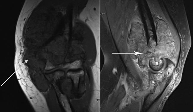
Magnetic resonance imaging (MRI) scan. MRI is the best imaging test to evaluate for the presence and extent of TGCT. MRI provides clear images of the body's soft tissues and joints.
- In localized TGCT, an MRI will show a nodular mass.
- In diffuse TGCT, an MRI will show extensive thickening of the joint lining throughout most of the joint or an extensive mass around tendons. For tumors inside of joints, there will possibly be bone changes and cartilage damage.
MRI can also help to identify blood within the tumor or joint, helping to make the diagnosis.
Positron emission tomography (PET) scans. While the contrast (dye) used in PET scans can show areas of TGCT, there is not sufficient data to support the routine use of PET scans in diagnosing this tumor.
Joint aspiration. In this procedure, fluid is removed from the joint with a syringe and analyzed. In many cases of TGCT, the joint fluid is bloody. If blood is found in a joint without a history of significant injury, TGCT should be suspected.
The diagnosis of TGCT can usually be made by your doctor based on your symptoms and what is seen on the MRI. In some cases, however, a biopsy may be needed to help confirm the diagnosis.
Treatment for Tenosynovial Giant Cell Tumor
As with all non-cancerous conditions, treatment decisions need to balance the risk of the condition with the potential benefits and complications of treatment. For patients without symptoms who do not want treatment, the tumor can be closely followed with assessments of pain and function, physical exams, and routine MRIs.
Because TGCT more commonly damages healthy tendons, cartilage, and bone and can grow to a large size, treatment typically involves surgery to remove the tumor and the damaged portions of the joint lining.
Localized TGCT Outside of a Joint
When localized TGCT involves a tendon, your surgeon will perform a procedure to remove the entire tumor. If the tumor has also damaged the tendon, your surgeon may repair or reconstruct it during the procedure to remove the tumor. Your doctor will discuss the various surgical options with you.
TGCT Inside a Joint
Surgical Procedures
When TGCT involves the inside of a joint, the options for specific surgical procedures depend on the joint involved, as well as the size and location of the tumor.
Arthroscopy. The tumor and damaged joint lining can be removed arthroscopically. This type of surgery is more often used for localized TGCT but can also be used for more diffuse disease. The risk of TGCT coming back is higher with arthroscopy than open surgery, especially for diffuse disease, but there are fewer potential complications with arthroscopy.
During arthroscopy the surgeon makes a few small incisions around the joint and inserts a small camera called an arthroscope. The camera shows pictures on a video monitor, and the surgeon uses these images to guide small surgical instruments to remove the tumor and the damaged synovium (joint lining tissue).
Open surgery. Diffuse TGCT requires removal of the entire joint lining to reduce the chances of the tumor returning. In many cases, this is best achieved with traditional "open" surgery.
Open surgery requires larger incisions than are used in arthroscopy and has a higher risk of complications. Benefits of open surgery include:
- It gives the surgeon full access to the joint.
- It enables the surgeon to safely and effectively remove both the mass and the joint lining.
- There is a lower risk of the tumor coming back.
Combined arthroscopic and open surgery. This approach can be used with diffuse TGCT of the knee.
- The surgeon uses open surgery to remove both the mass and the joint lining in the back of the knee.
- The surgeon uses arthroscopy to remove the tumor and joint lining in the front of the knee.
This combined method decreases the scale of the surgery, allowing for easier recovery.
Total joint replacement. In its end stages, widespread TGCT within joints can cause extensive destruction. Once the joint has become significantly damaged, sometimes the best option to improve pain and function may be a total joint replacement.
Total joint replacement is a procedure in which parts of a damaged joint are removed and replaced with metal and plastic parts. However, in some cases, the tumor may return even after the joint has been replaced.
Radiation Therapy
Radiation therapy can shrink tumors and is sometimes used to treat diffuse TGCT, especially for patients whose tumors have come back despite optimal prior treatment. However, there is not enough data on the use of radiation therapy for this tumor to support doctors reliably recommending it.
Radiation therapy is given via an external beam that is directed from outside the skin to the inside of the affected joint. While it can prevent additional recurrences, this method can also cause complications, including:
- Scarring and stiffness in and around the joint
- Damage to articular cartilage and bone
- The risk of a future radiation-induced cancer
Drug Therapy
There are currently two medications (Pexidartinib and Vimseltinib) available and several others being studied for the treatment of TGCT.
Pexidartinib and Vimseltinib specifically target the CSF-1R pathway, impacting the inflammation produced by TGCT. These medications can be considered for patients with symptomatic TGCT:
- Who are not candidates for surgery because of the size or location of the tumor
- For whom surgery could lead to significant change in or loss of function and affect quality of life
As with all medications, there are risks in using these drugs. Your doctor will talk to you about whether you are a good candidate for drug therapy and balance the potential benefits and risks with those of surgery.
Recovery After Surgery for TGCT
After surgery, physical therapy will be very important in helping you return to your daily activities. Specific exercises will help you regain strength and range of motion in the affected joint.
- Recovery from arthroscopic surgery usually requires a short course of physical therapy, after which you may return to normal activity.
- Open surgery is more extensive, so there is an increased risk of post-operative stiffness. A more involved and longer physical therapy program is often required for patients recovering from open surgery to treat diffuse TGCT. In this case, the return to all usual activities will take longer — possibly several months.
Because of the damage caused by the tumor, patients with TGCT — especially the diffuse form within joints — can have long-term issues with stiffness and pain and may need input from specialists in chronic pain management.
Localized TGCT rarely comes back after surgery, and patients usually need only limited follow-up.
Because of the higher recurrence rates for diffuse TGCT, patients with this form of the disease require follow-up with their doctor for several years after surgery. During these visits, your doctor may order tests such as an MRI — usually every 6 to 12 months — to check for recurrence.
Contributed and/or Updated by
Peer-Reviewed by
AAOS does not endorse any treatments, procedures, products, or physicians referenced herein. This information is provided as an educational service and is not intended to serve as medical advice. Anyone seeking specific orthopaedic advice or assistance should consult his or her orthopaedic surgeon, or locate one in your area through the AAOS Find an Orthopaedist program on this website.
The content on this website may have been translated from English into other languages using artificial intelligence tools. The translations are provided for the convenience of readers and may not been reviewed or verified by medical professionals. AAOS makes no guarantees regarding the accuracy, reliability, or completeness of the translated information and disclaims all liability for any issues arising from its use. Users are strongly encouraged to refer to the original English content and consult qualified healthcare professionals before making any medical decisions. By using the translated content, you acknowledge and accept these limitations and assume full responsibility for any reliance on the information provided.