






Diseases & Conditions
Synovial Chondromatosis
Synovial chondromatosis (at times also called synovial osteochondromatosis) is a benign condition that involves the synovium, which is the thin layer of tissue that lines the joints.
In synovial chondromatosis, cartilage and sometimes bone nodules occur inside the joint. There are two types of synovial chondromatosis:
- Secondary synovial chondromatosis occurs due to joint damage, such as trauma or arthritis. It is not a tumor. Treatment of secondary synovial chondromatosis depends on the underlying cause.
- Primary synovial chondromatosis is a rare noncancerous tumor and will be the focus of this article.
Primary synovial chondromatosis can affect any joint in the body but most commonly occurs in the knee. It is extremely unusual for synovial chondromatosis to affect more than one joint, and this tumor will not spread. Synovium also lines tendons outside of joints, so synovial chondromatosis sometimes occurs outside of a joint.
Although the condition is not cancerous, it can be aggressive and severely damage the affected joint, eventually leading to osteoarthritis. Early treatment is important to help relieve painful symptoms and prevent further damage to the joint.
Anatomy
The ends of bones that make up a joint, like the bottom of the thigh bone and top part of the shin bone in your knee, are covered with articular cartilage. Articuliar cartilage is a smooth substance that helps the bones glide over each other during movement.
Osteoarthritis occurs when the articular cartilage starts to break down and becomes worn and frayed. Moving the bones along this exposed joint surface is painful.
Thick bands of tissue surround the joint, forming a capsule that holds it together. The underside of the joint capsule, as well as ligaments within joints, are lined by a thin membrane called the synovium. Cells in the synovium produce a fluid that lubricates the joint.
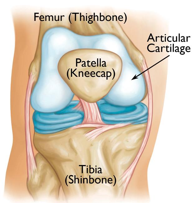
Description
In synovial chondromatosis, the cells in the synovium change into cells that can produce cartilage. Eventually, the cells form nodules of cartilage within the synovium. These nodules can sometimes break off from the synovium and become loose inside the joint.
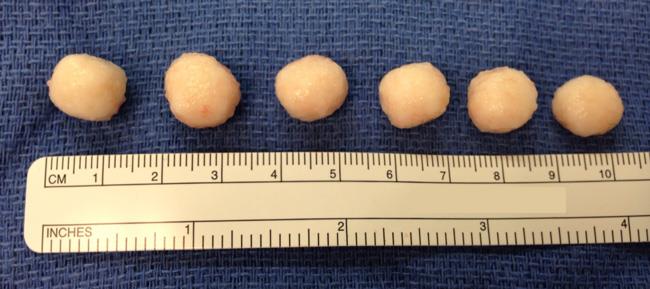
The size of the cartilage bodies inside the joint can vary — from the size of a small pill (or even smaller) to the size of a marble.
At first, these cartilage bodies are only within the synovium. However, the cartilage bodies may grow, calcify (harden), or ossify (turn into bone). Eventually, they can break free from the synovium and can then move around freely inside the joint space.
As they move around, the loose bodies can damage the smooth articular cartilage that covers the joint, causing osteoarthritis. Even bodies still within the synovium can damage the nearby articular cartilage. The bodies can also get trapped between bones that make up a joint. When this happens, the joint feels like it is getting stuck or it doesn't move like it used to.
In severe cases of synovial chondromatosis, the loose bodies may grow large enough to take up the entire joint space or enter into nearby tissues.
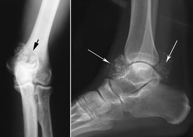
Synovial chondromatosis most often occurs in the knee, followed by the hip, shoulder, elbow, and ankle.
The condition most often occurs in patients between the ages of 30 and 50. Men are affected more than women.
Causes of Synovial Chondromatosis
The cause of synovial chondromatosis is unknown. The condition occurs spontaneously, meaning without a clear reason or trigger.
Various changes in genes have been found in synovial chondromatosis, confirming that it is a tumor. However, these gene changes are not specific to synovial chondromatosis, no consistent gene change has been identified, and the condition is not inherited.
Symptoms of Synovial Chondromatosis
The most common symptoms of synovial chondromatosis are similar to those of osteoarthritis. These include:
- Joint pain
- Joint swelling
- Limited range of motion in the affected joint (stiffness)
Other signs and symptoms may include:
- Fluid in the joint
- Tenderness
- A creaking, grinding, or popping noise during movement (crepitus)
- Locking
- Giving way
The nodules can sometimes be felt in joints that are close to the skin, such as the knee, ankle, and elbow joints. You may also be able to feel the nodules if the tumor involves tendons around the joint.
Diagnosing Synovial Chondromatosis
It is important to seek treatment for synovial chondromatosis as early as possible to help relieve painful symptoms and prevent the progression of osteoarthritis in the joint.
Physical Examination
Your doctor will talk to you about your general health and medical history and ask about your symptoms. They will then carefully examine the affected joint, looking for:
- Swelling
- Tenderness
- Limited range of motion
- Creaking or grinding noises during movement, an indication of bone-on-bone friction
Tests
Your doctor will order imaging tests to help diagnose synovial chondromatosis. Imaging tests will also help your doctor distinguish synovial chondromatosis from osteoarthritis. Sometimes, if primary synovial chondromatosis has caused significant osteoarthritis, it is hard to tell which came first (whether you have primary or secondary synovial chondromatosis).
X-rays. X-rays provide images of bone. Larger loose bodies are usually calcified or ossified and can be seen on X-ray. Smaller bodies and those that are not calcified or ossified may not show up.
Other imaging tests. Your doctor may order a magnetic resonance imaging (MRI) scan and/or computed tomography (CT) scan to better evaluate the joint and see where the synovium is involved. Cartilage bodies can typically be seen on both MRI and CT scans even when they are too small to see in X-rays.
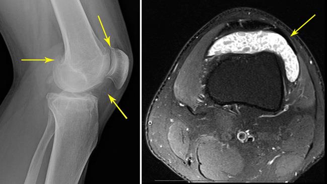
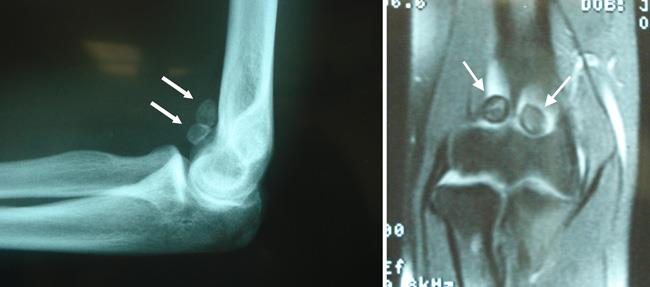
In addition to showing the loose bodies, imaging tests can also help your doctor determine whether you have any additional problems, such as fluid in the joint or signs of osteoarthritis (narrowing of the joint space and bone spurs).
Treatment of Synovial Chondromatosis
Nonsurgical Treatment
Observation. Depending on your symptoms, simple observation can sometimes be a treatment option. Your doctor will carefully consider a number of factors in determining whether observation is appropriate in your case.
During this time, your doctor will closely monitor the affected joint to check for the progression of osteoarthritis.
Surgical Treatment
Treatment for synovial chondromatosis typically involves surgery to remove the cartilage bodies, whether the bodies are loose or still within the synovium. Currently, there are no other treatment options (e.g., medications) for synovial chondromatosis.
Surgery can include just removing the cartilage or bone nodules or removing them along with some or all of the synovium (partial or complete synovectomy). The risk of the tumor coming back may be higher if only the loose bodies are removed. This is because the remaining synovium can continue to produce new cartilage bodies. However, it is unclear which procedure will lower the risk of the tumor coming back in which patients.
Surgery can be done using either an open procedure or an arthroscopic procedure. The technique your doctor uses will depend upon several factors, including:
- The number and location of the bodies
- The size of the bodies
- The condition of the synovium and how extensive the tumor is within the synovium
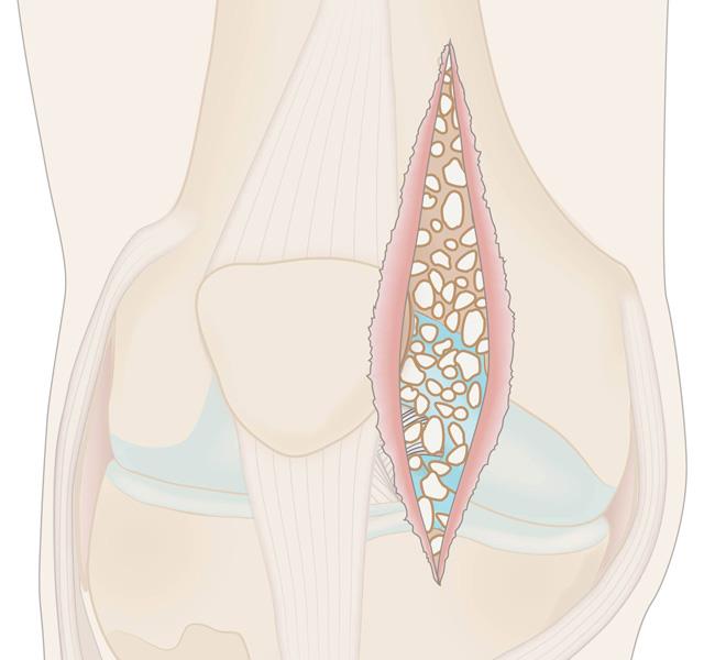
In a traditional open procedure, the doctor will usually make one or two large incisions.
In an arthroscopic procedure, the doctor will make smaller incisions and use miniature surgical tools to remove the loose bodies. Sometimes, your doctor may recommend using a combination of arthroscopic and open procedures to remove the tumor.
The end results of both open and arthroscopic procedures may be the same. The risk of the tumor coming back depends, in part, on how much the synovium was involved and in which parts of the joint. Your doctor will talk to you about which surgical technique is best in your case.
Recovery from Surgery
How long it takes to return to your daily activities depends on the type of procedure you have and which joint is involved. Your doctor will provide you with specific instructions to guide your rehabilitation.
Synovial chondromatosis may return in up to 25% of patients. For a period of time after surgery, your doctor will schedule regular follow-up visits to check for any recurrence.
Your doctor will also monitor the joint for any progression of osteoarthritis. The amount of damage synovial chondromatosis has already done to the joint will influence your chance of developing symptomatic osteoarthritis.
Contributed and/or Updated by
Peer-Reviewed by
AAOS does not endorse any treatments, procedures, products, or physicians referenced herein. This information is provided as an educational service and is not intended to serve as medical advice. Anyone seeking specific orthopaedic advice or assistance should consult his or her orthopaedic surgeon, or locate one in your area through the AAOS Find an Orthopaedist program on this website.
The content on this website may have been translated from English into other languages using artificial intelligence tools. The translations are provided for the convenience of readers and may not been reviewed or verified by medical professionals. AAOS makes no guarantees regarding the accuracy, reliability, or completeness of the translated information and disclaims all liability for any issues arising from its use. Users are strongly encouraged to refer to the original English content and consult qualified healthcare professionals before making any medical decisions. By using the translated content, you acknowledge and accept these limitations and assume full responsibility for any reliance on the information provided.