






Diseases & Conditions
Osteofibrous Dysplasia and Adamantinoma
Osteofibrous dysplasia (OFD) and adamantinoma are rare bone tumors that are not completely understood. They are most often found in the tibia (shinbone) but can occur in other bones.
- Osteofibrous dysplasia is a benign (noncancerous) tumor that typically develops during childhood. It does not spread to other parts of the body. Many cases are treated conservatively with careful observation over time.
- An adamantinoma is a malignant (cancerous) tumor that is capable of spreading and requires surgery to remove. It usually appears in adolescents and young adults after the bones have stopped growing.
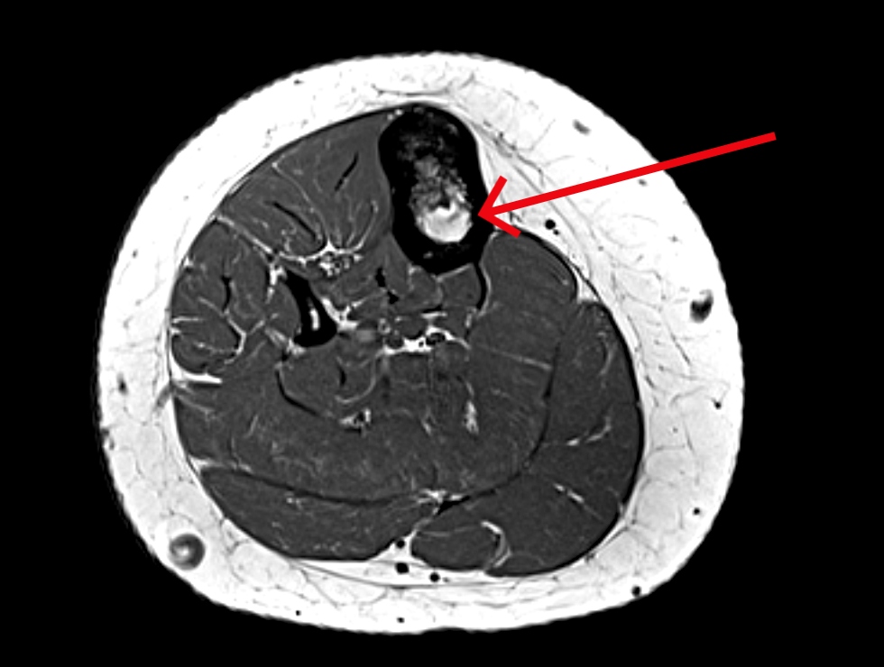
Doctors and researchers believe that OFD and adamantinoma might be related based on several similarities between the tumors, including location and appearance in X-ray images and under a microscope.
In addition, doctors have identified a third similar tumor — an OFD-like adamantinoma (OFDLA) — that includes a mix of cancerous and noncancerous tumor cells but is not known to spread to other parts of the body.
Although these three tumors may occur along a range of disease — with OFD on the benign end, adamantinoma on the malignant end, and OFD-like adamantinoma in the middle — it does not mean that they are different forms of the same tumor. Existing evidence does not show that OFD can progress to adamantinoma.
In addition, while these tumors can look similar under a microscope, more recent research has shown different gene changes in each, which means these tumors are distinct.
Description
Osteofibrous Dysplasia (OFD)
Osteofibrous dysplasia makes up less than 1% of all tumors that originate in bone. It usually develops in people younger than 20 years of age, most often occurring before the age of 10. Cases in people older than 20 can happen but are extremely rare.
OFD is slightly more common among males than females and appears at a somewhat younger age in boys.
Almost all OFD tumors occur in the long middle part of the tibia, with a few, rare reports of occurrences in the fibula (the smaller bone in the calf), as well as the long bones in the arm (humerus, radius, and ulna). OFD grows within the hard, cortical bone that makes up the outer portion of these bones and can cause the tibia to bend (bow).
Adamantinoma
Like OFD, adamantinoma is very rare. Fewer than 1% of all cancerous tumors that begin in the bone are adamantinomas.
These slow-growing tumors most often occur in the middle part of the tibia, and in many cases, the fibula is also affected. Although uncommon, adamantinomas have also been found in the bones of the arm, rib, pelvis, foot, and spine.
Like OFD, adamantinoma tumors start in the hard, cortical bone, but they are more aggressive. They can destroy the outer cortex or spread into the central canal of the bone. In many cases, adamantinomas also affect the muscles and other soft tissues around the bone.

Unlike OFD, in about 15% of cases, adamantinomas metastasize (spread) to other areas of the body — most often the lungs, lymph nodes, and other bones. There have been reports of metastasis occurring years after the initial diagnosis and subsequent treatment.
Although adamantinomas can develop in anyone at any age, they tend to occur in people older than those with OFD. They often appear between the ages of 20 and 50, and most occur between the ages of 20 and 30. These tumors are slightly more common in men than in women.
Cause of OFD and Adamantinoma
The cause of OFD and adamantinoma is unknown. There has been no proven connection between the development of these bone tumors and exposure to chemicals, radiation, or any particular activities.
Symptoms of OFD and Adamantinoma
In many cases of both OFD and adamantinoma, patients do not have symptoms, and the condition is discovered in an X-ray that is taken for another reason, such as an injury.
When symptoms do occur, they tend to vary from person to person. The most common symptoms of both OFD and adamantinoma include:
- Swelling over the tumor site
- Pain in the area of the tumor
- A break due to the tumor weakening the bone
- When the tumor develops in the tibia, the lower leg may be curved due to bowing of the bone
Diagnosing OFD and Adamantinoma
Medical History and Physical Examination
Your doctor will talk to you about your general health and your symptoms to get a good history of the problem. During the physical examination, your doctor will look for tenderness over the bone, swelling, or a mass in the area of your symptoms.
Tests
The signs and symptoms of OFD and adamantinoma are similar to many other types of bone tumor. Your doctor may use several tests to distinguish between different tumors and make an accurate diagnosis.
- X-rays. Because X-rays provide clear pictures of bone, they are very helpful in diagnosing bone tumors.
Different types of tumors look different on X-rays. For example, an OFD has very obvious borders and can look like one big tumor or several smaller areas of tumor. An adamantinoma has a "soap bubble" appearance in X-rays and tends to be larger than an OFD.

- Other imaging scans. You may need more imaging tests to further evaluate these tumors. Your doctor may also request computerized tomography (CT) scans and magnetic resonance imaging (MRI) scans to help further define the tumor. These scans can provide more detail, especially of soft tissues. They can also provide cross-sectional images.
A CT scan or MRI scan will show your doctor more precisely where the tumor is located and what its specific characteristics are. For example, in a CT or MRI scan, your doctor can see if the adamantinoma tumor has broken through an area of the bone into the middle (canal) of the bone or into the muscles on the outside of the bone.
Biopsy. A biopsy may be needed to confirm whether the tumor is OFD, an adamantinoma, or an OFD-like adamantinoma. In a biopsy, a tissue sample of the tumor is taken and examined under a microscope. Your doctor or a radiologist may give you a local anesthetic to numb the area and take a sample using a needle. Biopsies can also be performed as a small operation.
If your doctor diagnoses the tumor as adamantinoma, you may require some additional tests, such as a CT scan of your chest or a bone scan, to determine whether the disease has spread to other areas.
Treatment of OFD and Adamantinoma
Nonsurgical Treatment
Because OFD is not cancerous, treatment typically involves observation. Your doctor will monitor the tumor with regular X-rays taken every few months to make sure the tumor is not getting bigger. An OFD-like adamantinoma is treated the same way, although there is not always agreement on treatment of OFDLA
If the tumor is weakening the bone enough to cause it to bow or change shape, your doctor may recommend wearing a brace. This may also prevent the tumor from causing the bone to break.
Surgical Treatment of OFD and OFDLA
Surgery may be recommended to remove the tumor and stabilize the bone if an OFD or OFD-like adamantinoma causes:
- Pain
- Serious bone deformity
- Fractures (or a bone that is at risk of breaking because of the amount of bowing)
Surgery may also be recommended for large OFDLA tumors.
Surgery for OFD and OFDLA includes either:
- Curettage (scraping out of the tumor) and bone grafting
- Removing the part of bone involved with the tumor
Curettage is more likely to be recommended for OFD than removal of the bone. Curettage is a less aggressive surgery than removing the involved part of bone, but there may be a somewhat higher rate of the tumor coming back.
Surgical Treatment of Adamantinoma
Adamantinomas always require surgery to remove the area of bone and any surrounding muscle involved with the tumor. They do not respond to other cancer treatments, such as chemotherapy and radiation.
- Limb salvage surgery. In this procedure, your doctor removes the section of bone where the tumor is located, as well as some healthy tissue surrounding it. To perform limb salvage surgery, there needs to be enough bone and muscle remaining after the tumor is removed to make the limb useful. The hole where the bone is removed is filled with a bone graft to stabilize the bone and promote healing with new bone growth. Bone graft is bone taken from a cadaver (allograft) or from another bone in your body (autograft). Sometimes, the hole requires reconstruction with a metallic implant to stabilize the bone.
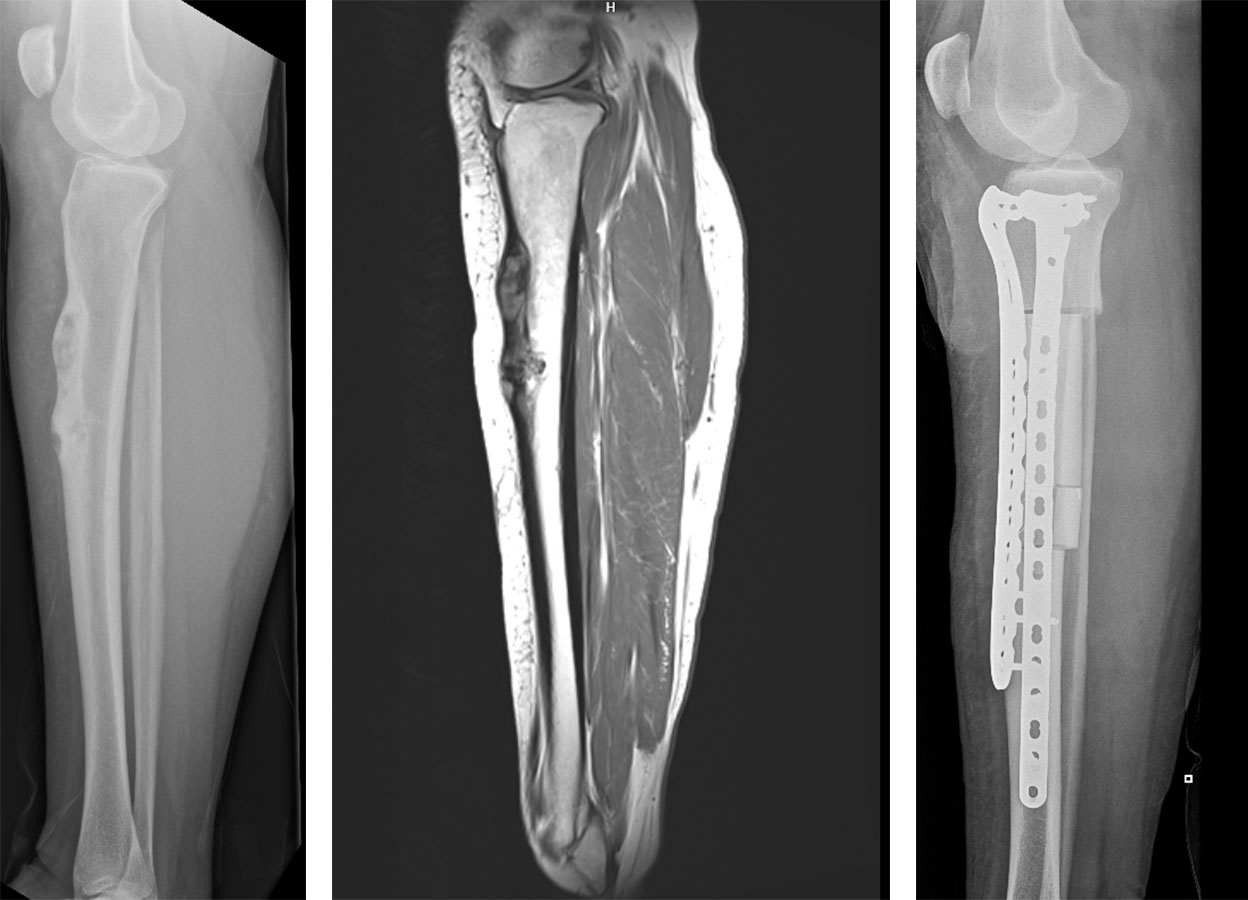
- Amputation. All or part of the limb may need to be removed if the adamantinoma cannot be completely removed with limb salvage surgery, or if important nerves or blood vessels are involved. Your surgeon will talk to you about the benefits of limb salvage versus amputation.
How long it takes to return to daily activities depends on how large the tumor was, where it was located, and what kind of surgery you had. Your doctor will provide you with specific instructions to guide your rehabilitation.
You will need regular doctor visits and tests every few months for several years after surgery to find out if the tumor has come back.
Summary
- An OFD is a benign tumor that has not been shown to become cancerous.
- Adamantinomas are cancerous and may come back in some people, sometimes many years later. Because of this risk of recurrence, you will need long-term monitoring by your doctor.
- While OFD and adamantinoma are thought to potentially be related, there is no good evidence that an OFD turns into an adamantinoma.
Contributed and/or Updated by
Peer-Reviewed by
AAOS does not endorse any treatments, procedures, products, or physicians referenced herein. This information is provided as an educational service and is not intended to serve as medical advice. Anyone seeking specific orthopaedic advice or assistance should consult his or her orthopaedic surgeon, or locate one in your area through the AAOS Find an Orthopaedist program on this website.
The content on this website may have been translated from English into other languages using artificial intelligence tools. The translations are provided for the convenience of readers and may not been reviewed or verified by medical professionals. AAOS makes no guarantees regarding the accuracy, reliability, or completeness of the translated information and disclaims all liability for any issues arising from its use. Users are strongly encouraged to refer to the original English content and consult qualified healthcare professionals before making any medical decisions. By using the translated content, you acknowledge and accept these limitations and assume full responsibility for any reliance on the information provided.