






Diseases & Conditions
Giant Cell Tumor of Bone
A giant cell tumor of bone is a rare, usually benign (noncancerous) bone tumor. Although it is not considered cancer, it can behave aggressively by growing into and destroying nearby bone. As the tumor grows it can weaken the bone, increasing the risk of fracture and often affecting the function of the nearby joint.
Giant cell tumors most commonly develop at the end of long bones near joints after skeletal growth is complete. They are most often found around the knee, especially in the lower end of the femur (thigh bone) and the upper end of the tibia (shin bone).
Treatment usually involves surgery to remove the tumor while preserving as much healthy bone and joint function as possible. Depending on the size and location of the tumor, medications such as denosumab may be used before surgery to shrink the tumor or in cases where surgery is not possible.
Because giant cell tumors can come back after surgery, regular follow-up with imaging is important to monitor for recurrence, especially if you are treated with denosumab before surgery.
How and Where Do Giant Cell Tumors Form?
Many bone tumors and normal bone tissue contain types of giant cells. Giant cells in normal bone are called osteoclasts and are necessary to remove damaged bone. However, the giant cells seen in giant cell tumors are different than those seen in normal bone.
A giant cell tumor of bone is diagnosed based on its unique appearance under the microscope (see photo below). In addition to numerous large or "giant cells" that are formed by the fusion of several individual cells and contain multiple nuclei, the tumor also contains a background of abnormal stromal cells. These are the true tumor cells and help distinguish a giant cell tumor of bone from other bone conditions.
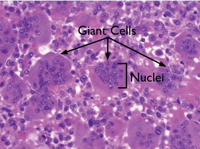
Another feature that makes giant cell tumors unique is where they develop.
- Most bone tumors begin in the metaphysis, the wider part of the bone near the end.
- In contrast, giant cell tumors almost always develop in the epiphysis, the end of the bone that forms part of a joint. This characteristic location, combined with its appearance on imaging and under the microscope, helps doctors make the diagnosis.
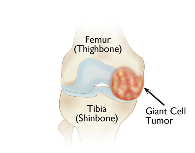
Giant cell tumors of bone most often develop near the knee, either in the lower end of the femur or the upper end of the tibia.
Other common locations include the:
- Wrist (lower end of the lower arm bone)
- Hip (upper end of the thighbone)
- Shoulder (upper end of the upper arm bone)
- Lower back (connection of the spine and pelvis)
In rare cases, a patient can have multiple giant cell tumors in different bones. This is called multi-centric giant cell tumor of bone.
Giant cell tumors are extremely rare, affecting approximately 1 person per million each year. Most giant cell tumors occur in patients between 20 and 40 years of age. They are uncommon in children or in adults over 65 years of age. They occur slightly more often in women than in men.
While giant cell tumors are typically benign (noncancerous), they can grow quickly and destroy bone close to a joint. In rare cases, they can spread to the lungs while still being benign. On very rare occasions, a giant cell tumor can be cancer.
Causes of Giant Cell Tumor of Bone
The exact cause is not known. These tumors develop on their own and are not linked to injury, diet, or environmental factors. They are also not inherited (passed down from a parent).
In some cases, a build-up of giant cells (osteoclasts), typically in multiple bones, may be associated with overactivity of the parathyroid glands (hyperparathyroidism). This collection of giant cells is not a tumor; it represents a separate medical condition and is not the same as having a giant cell tumor.
Symptoms of Giant Cell Tumor of Bone
- The most common symptom is pain in the area of the tumor. Pain may also occur with movement of the nearby joint. It often gets worse with activity and improves with rest. Pain is usually mild at first but gradually worsens as the tumor grows.
- You may also have swelling, stiffness, and decreased range of motion in the nearby joint.
- In some cases, the weakened bone may break, causing sudden and severe pain. This is called a pathologic fracture — a fracture caused by weakening of the bone from the tumor rather than a traumatic injury.
- Some patients may not have pain at all and may instead notice swelling or a lump.
Diagnosing Giant Cell Tumor of Bone
Your doctor will perform a thorough physical examination and use imaging tests, most often X-rays, to evaluate the area and diagnose a giant cell tumor.
Tests
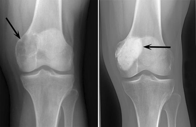
X-ray. X-rays provide clear images of bone. Your doctor will order an X-ray to help confirm the diagnosis.
On X-ray, a giant cell tumor appears as an area of destruction near the joint. The bone may look weakened or "eaten away." In some cases, a thin rim of remaining bone is seen around the tumor, and the bone may appear slightly expanded.
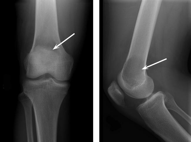
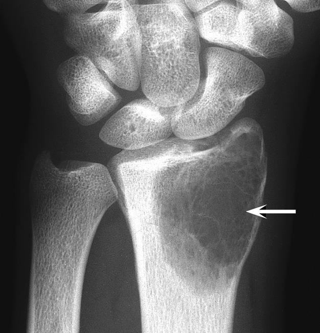
Other imaging tests. Your doctor may order a magnetic resonance imaging (MRI) scan to better evaluate the tumor and the area surrounding it, as giant cell tumors can break through the bone and grow in surrounding soft tissues.
A CT scan or chest X-ray may also be done at the time of the initial diagnosis to see if the tumor has spread to the lungs.
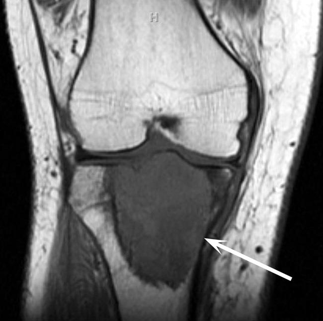
Biopsy. A biopsy is usually needed to confirm the diagnosis of a giant cell tumor, especially as the tumor can appear aggressive on X-rays or MRI. In a biopsy, a sample of tissue is taken from the tumor and examined under a microscope.
A biopsy can be performed under local anesthesia with a needle or as a small open operation.
Treatment of Giant Cell Tumor of Bone
Without treatment, a giant cell tumor will continue to grow and destroy the surrounding bone, so treatment is always necessary. As with all health conditions, it is important to talk to your doctor about the risks and benefits of available treatments.
The goals of treatment are:
- Remove the tumor
- Prevent damage to the bone and nearby joint
- Prevent the tumor from coming back
Surgery is the preferred treatment for giant cell tumors; however, there are some cases in which a tumor cannot be removed safely or completely through surgery. In this case, your doctor may recommend nonsurgical treatment.
Nonsurgical Treatment
Nonsurgical treatment may include:
Radiation. Radiation therapy may sometimes be used to shrink giant cell tumors in areas where surgery may be difficult to perform without damaging sensitive tissues — such as the spine. However, radiation therapy can result in the formation of cancer in some patients, so it is used only in rare cases.
Tumor embolization. During this procedure, specific arteries that supply blood to the tumor are blocked off. Without their supply of oxygen and nutrients, the tumor cells begin to die.
- Most often, embolization is performed before surgery to decrease the amount of blood lost during surgery.
- Embolization may also be used on its own in cases where surgery cannot be performed. However, because the body typically makes new blood vessels, the tumor can come back and continue to grow after embolization.
Medication. The FDA has approved the use of an injectable medication (denosumab) for the treatment of giant cell tumors. The medication is injected under the skin, not into the tumor. It works by targeting a special receptor on the giant cells. This decreases activity and slows down the breakdown of bone. However, because the tumor cells are located between the giant cells, the tumor will come back after stopping the treatment.
The medication is sometimes used in cases where surgery cannot be performed or for tumors that come back. It can also be used to decrease the size of larger tumors before surgery; however, data shows that the risk of the tumor coming back in the same spot may be higher in patients who are initially treated with denosumab.
Surgical Treatment
Surgery has proven to be the most effective treatment for giant cell tumors. Surgical treatment may include:
Curettage. Curettage is the surgical procedure most commonly used to treat giant cell tumors. In curettage, special instruments are used to scrape the tumor out of the bone.
Usually after curettage, chemicals such as liquid nitrogen or hydrogen peroxide are placed inside the bone cavity to try to kill any remaining tumor and keep the tumor from coming back. In some cases, an argon gas laser will be used to help reduce the risk of recurrence.
After curettage, the cavity is often filled with bone cement; this not only helps to kill any remaining tumor but also helps to stabilize the bone. Bone grafts can also be used to fill the bone cavity.
Other procedures. If a giant cell tumor has caused significant damage to bone or the nearby joint, more complex surgery may be needed. In these cases, your surgeon may remove the bone involved with the tumor and rebuild the affected area (often including the joint) using:
- Bone grafts (donor bone or your own bone)
- Artificial joints (joint replacement surgery)
- A combination of both
The goal of surgery is to restore strength and function so you can return to normal daily activities as much as possible.
In rare cases, a giant cell tumor may spread to the lungs. If the tumor spreads, surgical removal of the tumor in the bone and the affected area of the lung can often cure the disease. However, if the areas in the lung are small, your doctor may decide to monitor them closely. If the tumor continues to grow despite surgery, you may be a candidate for medical treatment.
Outcomes
Results after treatment depend on several factors, including:
- Age of the patient
- Size and location of the tumor
- Type of treatment used
Giant cell tumors can come back after treatment, so long-term follow-up is very important. Patients typically need regular check-ups for several years. These visits usually include X-rays of the original tumor site, as well as chest X-rays or CTs to monitor the lungs.
Contributed and/or Updated by
Peer-Reviewed by
AAOS does not endorse any treatments, procedures, products, or physicians referenced herein. This information is provided as an educational service and is not intended to serve as medical advice. Anyone seeking specific orthopaedic advice or assistance should consult his or her orthopaedic surgeon, or locate one in your area through the AAOS Find an Orthopaedist program on this website.
The content on this website may have been translated from English into other languages using artificial intelligence tools. The translations are provided for the convenience of readers and may not been reviewed or verified by medical professionals. AAOS makes no guarantees regarding the accuracy, reliability, or completeness of the translated information and disclaims all liability for any issues arising from its use. Users are strongly encouraged to refer to the original English content and consult qualified healthcare professionals before making any medical decisions. By using the translated content, you acknowledge and accept these limitations and assume full responsibility for any reliance on the information provided.