






Diseases & Conditions
Chondroblastoma
Chondroblastoma (kon-dro-BLAST-oma) is a rare type of benign (noncancerous) tumor that grows at the ends of the body's long bones, close to the joints. Fewer than 1% of all bone tumors are chondroblastomas.
Most often, the tumors develop at the ends of the femur (thighbone), upper end of the tibia (shinbone), or upper end of the humerus (upper arm bone). They typically occur in adolescents and young adults and are more common in males.
Although chondroblastomas are not cancerous, they can continue to grow if left untreated. A growing tumor can destroy the surrounding bone, making the bone painful or making it painful to move the nearby joint. The area around the tumor, including the nearby joint, can also become swollen.
Treatment for a chondroblastoma almost always involves surgery to remove the tumor and prevent damage to the bone near the affected joint.
Description
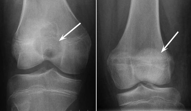
Most chondroblastomas are found close to the knee joint — either at the lower end of the femur (thighbone) or the upper end of the tibia (shinbone).
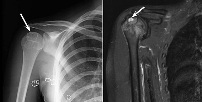
They are also found in the shoulder at the top of the humerus (upper arm bone). This type of chondroblastoma is often referred to as a Codman tumor.
Less common locations for chondroblastomas include the pelvis, hip, kneecap, and foot.

Chondroblastomas are made up of cells called chondroblasts. Chondroblasts are immature cartilage cells, which is how they got their name ("chondro" means cartilage and "blast" means embryo).
The tumors are thought to arise from the growth plates (physes) at the ends of the bones. This is where bones grow and become longer during childhood and adolescence. Chondroblastomas can also arise in smaller areas of growth centers (apophyses) that are on the surfaces of bones and that form areas of attachment of ligaments and tendons.
Chondroblastomas typically occur in people ages 10 to 25 years, with an average age of 19 to 23 years. They are twice as likely to develop in males than in females.
Chondroblastoma is one of the few benign bone tumors with the potential to spread, or metastasize, to the lungs. This happens in less than 2% of cases. Because of this extremely rare likelihood of spreading to the lungs, the World Health Organization (WHO) has changed their classification of this tumor from "rarely metastasizing" to completely benign.
Even though a more aggressive chondroblastoma may metastasize, it is still considered a benign tumor.
Causes of Chondroblastoma
The cause of chondroblastomas is not clear. .
While chondroblastomas occur near areas of growing cartilage, they do not produce the same type of normal cartilage that makes up the growth plates or that forms the protective surfaces within joints. More than 90% of chondroblastomas have a specific gene mutation which can lead to loss of control of the growth of cartilage cells.
There has been no proven connection between the development of the tumors and exposure to chemicals or radiation or any particular activities.
The risk of developing a chondroblastoma does not run in families and is random.
Symptoms of Chondroblastoma
Pain is the most common symptom of a chondroblastoma and often is present for a long time. Because the tumors are typically found near joints, it is joint pain and swelling that will often prompt a visit to the doctor. There may also be pain coming from the involved bone.
Over-the-counter pain medications such as acetaminophen and ibuprofen may help relieve this pain at first but will stop helping as the tumor grows in size.
Other signs and symptoms of a chondroblastoma may include:
- Joint stiffness
- Joint swelling
- Muscle atrophy (wasting away of the surrounding muscle from lack of use)
- A limp (when the tumor is in the lower extremities)
A chondroblastoma is usually small and contained within the bone, so patients do not normally see or feel a mass.
Diagnosing Chondroblastoma
Physical Examination
The doctor will talk to you about your or your child's medical history and general health and ask about symptoms. They may ask about symptoms such as fever or chills because pain or swelling in a joint in younger people can be related to an infection rather than a tumor.
The doctor will try to learn as much as possible about your or your child's pain. Pain at night or pain that does not go away with rest is usually more concerning. This is because pain caused by an injury will usually decrease when the injured area is not being used.The doctor will then examine the painful area, looking for:
- Tenderness over the bone
- Limited range of motion in the nearby joint
- Swelling around the joint
- Redness or warmth (to make sure the pain and swelling are not due to an infection)
The doctor will use imaging and other tests to help diagnose a chondroblastoma.
Tests
X-rays. X-rays provide images of bone and are very helpful in diagnosing tumors. However, they may not be able to definitively diagnose a chondroblastoma.
Most chondroblastomas are small (1 to 4 cm), round tumors within the ends of bones. They appear right next to joints or, sometimes, on the surface of bones. On X-ray images, they are often surrounded by a thin rim of bone that looks white on the X-rays.
Some chondroblastomas will cause the edge of the bone to push out. But the tumors rarely extend past the bone and into the surrounding soft tissue. In about 25 to 40% of cases, an X-ray will show calcifications (white spots) inside the tumor.
Computed tomography (CT) scans. More detailed than a plain X-ray, a CT scan can help the doctor further evaluate the tumor and plan your or your child's treatment. Calcifications inside the tumor are more likely to be visible on a CT scan.
Magnetic resonance imaging (MRI) scans. MRI scans provide clear images of the body's bone marrow and soft tissues. This imaging may be preferred over CT because of the radiation exposure you get with CT.
An MRI will help the doctor better see the edges of the tumor and whether it has expanded out of the bone and into nearby tissues. It will also show areas of swelling in the bone marrow and soft tissues that surround this tumor.
Biopsy. A biopsy is needed to confirm the diagnosis of chondroblastoma. In a biopsy, a sample of tissue is taken from the tumor and examined under a microscope.
A biopsy can be performed under local anesthesia with a needle. The other approach is to perform a small open biopsy in the operating room. A radiologist will often perform the needle biopsy, while your surgeon will perform the open biopsy.
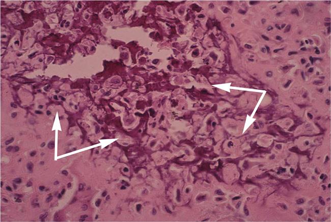
Under a microscope, chondroblastomas have a background that looks like cartilage and a mix of cells, some of which look like cartilage-making cells (these have nuclei that look like coffee beans). Calcifications may be seen weaving throughout the tumor in a pattern that resembles chicken wire.
Differential Diagnosis
A biopsy will help the doctor tell a chondroblastoma apart from other tumors that have a similar appearance, such as a giant cell tumor, enchondroma, aneurysmal bone cyst (ABC), infection of the bone (osteomyelitis), or chondroblastoma-like osteosarcoma.
- Like chondroblastomas, giant cell tumors also typically grow next to joints or in apophyses but typically occur in people between the ages of 20 and 40.
- Enchondromas can look similar to chondroblastoma on X-rays, as both tumors make cartilage; however, enchondromas usually don't cause symptoms, especially in younger people.
- ABCs occur in patients of the same age as those with chondroblastomas, and about 10 to 15% of chondroblastomas can contain areas of ABC.
It is important to tell a chondroblastoma apart from a more aggressive tumor, such as a chondroblastoma-like or giant cell-rich osteosarcoma. A pathologist (a doctor who identifies diseases by studying abnormal cells) will review the tissue sample and do special tests to help determine the diagnosis.
Treatment of Chondroblastoma
Without treatment, a chondroblastoma will likely continue to grow and damage the surrounding bone, so treatment is almost always necessary. The goals of treatment are to:
- Remove the tumor
- Prevent damage to the bone and joint
Surgery is often the preferred treatment for chondroblastomas; however, there are some cases in which a tumor cannot be removed safely or effectively due to its location or size. In this case, the doctor may recommend nonsurgical treatment.
Nonsurgical Treatment
Nonsurgical treatment may include:
Radiofrequency ablation (RFA). In this procedure, the tumor is heated and destroyed with a high-frequency electrical current.
This procedure works best for small lesions. It can also be used when more extensive surgery might lead to unacceptable complications, especially if the tumor is in an area that is challenging to access with surgery, such as the hip.
However, RFA can damage the cells in the nearby growth plate or joint. It is important to discuss the pros and cons of each treatment option with your doctor.
Surgical Treatment
Most often, surgery is used to treat chondroblastomas. Surgical treatment may include:
Curettage. This is the procedure most commonly used to treat a chondroblastoma. In curettage, special instruments are used to scrape the tumor out of the bone. Once a chondroblastoma is curetted, it is unusual for it to return.
Curettage can be supplemented with other treatments, such as use of a burr to make sure all of the tumor has been removed or chemicals placed within the bone to destroy any remaining tumor cells. Chemicals that can be used include hydrogen peroxide or liquid nitrogen. However, studies do not indicate which additional treatment works best. Your doctor will discuss this with you. Once a chondroblastoma is curetted, it is unusual for it to return.
Chondroblastomas can be difficult to treat, as they are near growth plates and joints. While surgeons are very careful during surgery, these structures can be damaged by curettage, use of a burr, or use of chemicals, especially the extreme cold from liquid nitrogen. To avoid causing damage, surgeons may be less aggressive with treatment, increasing the likelihood that the tumor will come back and need additional treatment.
After curettage, the doctor may fill the cavity with a bone graft to help stabilize the bone. A bone graft is taken either from a donor (allograft) or from another bone in your body (autograft), most often the pelvis. The doctor may also use a bone cement mixture to fill the hole. While bone cement may decrease the risk of recurrence and help stabilize the bone, there is risk of damage to the nearby growth plate.
Resection. Depending on the location and size of the tumor, the doctor may remove the entire section of bone containing the tumor, rather than performing curettage. This requires reconstructing the bone and often the nearby joint.
In rare cases, a chondroblastoma can spread, most often to the lungs. However, the tumor that spreads still looks benign under the microscope. If the tumor spreads, surgical removal of the metastasis is necessary. This will typically result in a cure.
Studies of the use of the drug denosumab to treat metastasis from chondroblastoma are underway.
Surgical Complications
The most serious complication of surgical treatment is the tumor coming back. Other potential complications include:
- Infection.
- Fracture or collapse of the bone through the treated area, though this is rare.
- Damage to the growth plate causing abnormal bone growth (shortening or angulation of the arm or leg). This damage can be related to the tumor invading the growth plate or injuries to the growth plate during curettage or other treatments.
Outcomes
Your or your child's outcome after treatment will depend on a number of factors, including:
- Age and overall health
- The size and location of the tumor
- The method of treatment
Even with successful surgery, a chondroblastoma can damage the normal cartilage that surrounds and protects the joints, so some patients may develop arthritis over time.
Once chondroblastoma is treated with curettage, the risk of it coming back is about 10 to 20%. The patient's age and the location of the tumor are factors that increase the risk of the tumor coming back. The younger the patient, the more likely they are to have a recurrence.
If a tumor does come back, it usually does so within a few months to a few years. For this reason, the doctor will monitor you or your child with regular check-ups and X-rays for at least a few years. If a tumor recurs, it can be treated using the same methods. The doctor will talk to you about all of your options.
Contributed and/or Updated by
Peer-Reviewed by
AAOS does not endorse any treatments, procedures, products, or physicians referenced herein. This information is provided as an educational service and is not intended to serve as medical advice. Anyone seeking specific orthopaedic advice or assistance should consult his or her orthopaedic surgeon, or locate one in your area through the AAOS Find an Orthopaedist program on this website.
The content on this website may have been translated from English into other languages using artificial intelligence tools. The translations are provided for the convenience of readers and may not been reviewed or verified by medical professionals. AAOS makes no guarantees regarding the accuracy, reliability, or completeness of the translated information and disclaims all liability for any issues arising from its use. Users are strongly encouraged to refer to the original English content and consult qualified healthcare professionals before making any medical decisions. By using the translated content, you acknowledge and accept these limitations and assume full responsibility for any reliance on the information provided.