






Diseases & Conditions
Spinal Deformity in Children with Myelomeningocele
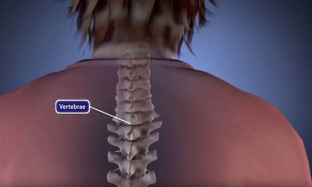
Myelomeningocele, or spina bifida cystica, is a birth defect. The defect is there when the child is born. It develops during the early weeks of fetal development. It is the most common type of spina bifida. It is also the most serious type. Spina bifida is a neural tube defect. According to the Centers for Disease Control, it affects about 1,400 babies born each year.
In babies with myelomeningocele, some bones of the back do not form in the usual way. These bones are called vertebrae. They do not close fully around the spinal cord. The bones leave a gap around the cord. As a result, parts of the spinal cord, nerves, spinal fluid, and nearby tissues push through the open backbone. These tissues are exposed. They often form a sac, which sticks out from the baby's back.
The opening or sac can form anywhere along the back. It most often forms in the lower back. Infants often lose nerve function below the level of the defect in the spine. The site of the defect affects which nerves work. Many infants and children have loss of feeling, paralysis, and bowel and bladder problems. This can change how the child feels and moves. The paralysis can cause deformities of the legs, feet, and back.
Many children with myelomeningocele develop severe spinal deformities, such as scoliosis. This article focuses on treatment for spinal deformities linked to myelomeningocele.
Children with myelomeningocele may have many medical problems. Treatment involves a team of doctors and health professionals from different medical specialties working together to provide care.
What Causes Myelomeningocele?
The cause of myelomeningocele is unknown.
Taking folic acid lowers the risk of neural tube defects, such as spina bifida. Folic acid helps lower this risk. These defects form very early in pregnancy. Doctors advise women who may become pregnant to take folic acid before pregnancy. They should also take it during pregnancy. As a result, the rate of neural tube defects, including myelomeningocele, is falling.
A spinal problem, or curvature, is common in more severe cases of myelomeningocele. The curvature may be caused by:
- Weak trunk muscles that allow the spine to collapse (neuromuscular); the weak muscles do not hold the spine straight
- Vertebrae that formed in an abnormal way (congenital); the bones have an unusual shape from birth
- A combination of both causes
The three most common types of spinal deformity are listed below. Each type changes the shape of the back:
- Scoliosis (a sideways curve of the spine to the left or right). In children with myelomeningocele, scoliosis often results from poor neuromuscular control. Weak trunk muscles cannot support the spine. This type of scoliosis may be linked to tethering of the spinal cord.

- Kyphosis (an exaggerated round back). Kyphosis can form because paralysis causes an imbalance in a child's trunk muscles. A very severe form is called myelokyphosis. It is very stiff. It may also cause skin breakdown over the apex, or highest point, of the prominence.
- Lordosis (an exaggerated sway back). Lordosis can occur when a child's hip muscles are tight. The tight muscles push the pelvis forward. This causes an exaggerated swayback.
Doctor Examination
A child with myelomeningocele needs ongoing care from several medical specialists. Each doctor treats one of the child's health issues.
- An orthopaedic surgeon treats bone or muscle problems that limit the child's ability to function
- A neurosurgeon checks the health of the child's brain and spinal cord
- A urologist treats bowel and bladder problems
- A physical rehabilitation doctor (physiatrist) helps customize braces and make any needed wheelchair changes
A pediatric orthopaedic surgeon leads treatment for a spinal curvature. However, these children have a wide range of medical needs. The whole medical team must take part. The team shares plans and goals for the child. This helps the child have the best outcome.
Physical Examination
Most children with myelomeningocele use wheelchairs because of partial or complete paralysis in their legs. A spinal curvature can cause poor sitting balance. This may place too much pressure on some areas and cause skin problems. For example, the kyphosis prominence may press against the back of the wheelchair. This can cause pressure sores that are difficult to heal.
A kyphosis curve may also force a child to lean forward. The child may need to use their hands for constant support. Without the use of their hands, the child cannot perform many daily activities.
The orthopaedic surgeon will examine the child's spinal curve. The doctor will also check sitting balance, skin breakdown, and daily function. The exam shows how the curve affects the child.
Imaging Tests
X-rays. X-rays of the spine help the doctor check the condition and position of the child's bones.
An X-ray will also show the doctor how severe the curve is. The doctor measures the degree of the curve on the X-ray. This helps the doctor choose the right treatment.
In general, curves of 20° or more are serious and need treatment. Curves greater than 45° to 50° will likely need surgery to restore a more normal posture.
Magnetic resonance imaging (MRI) scans. MRI scans give clear images of soft tissues. They help the doctor evaluate the health of the spinal cord. An MRI scan does not use X-rays.
Treatment
Many spinal deformities in children with myelomeningocele get worse over time. As the spinal curve grows, the risk of skin and functional problems rises. Severe curves can also reduce the space in the chest. This may cause heart problems. It may also cause lung problems.
Nonsurgical Treatment
Bracing. The doctor may advise a brace if the child is still growing and the spinal curve is flexible. A brace cannot straighten a spinal curve. However, it may slow the curve as it gets worse. This may allow the child to grow more before surgery is needed.
Each brace must be custom-made. The brace must fit well at all times. It should not cause skin irritation or other problems. The team monitors the skin as the child grows.
Wheelchair modification. A shifted pelvis may affect a child's balance. Changes to the wheelchair can improve posture. Like a brace, these changes may improve function and slow the curve as it gets worse. However, wheelchair modification is not a cure.
Surgical Treatment
Surgery may be needed if a brace or wheelchair changes do not control the spinal curve as it gets worse. Surgery may also be needed if the child first comes to the doctor with a severe spinal curve.
The operation used to treat a spinal curve is a spinal fusion. The surgeon realigns and joins the curved vertebrae. The vertebrae then heal into one solid bone. The goal is one strong and stable part of the spine. Screws, rods, hooks, or wires hold the bones in place. Spinal fusion can:
- Straighten the spine to improve posture, so the child does not need to use their hands and arms to support the upper body
- Prevent pressure points caused by uneven sitting, which can make the skin break down
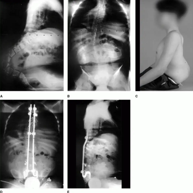
Surgery for spinal deformities linked to myelomeningocele can be difficult because of:
- A higher risk of surgical complications
- Other medical problems, such as hydrocephalus, skin conditions, and urinary tract infections
However, careful preoperative planning can lead to successful outcomes. This planning involves a team of medical professionals. The team makes its plan before the child has surgery. The plan must meet all the child's health needs. The team will watch the child as they grow. New fusion implant designs may also help.

Contributed and/or Updated by
AAOS does not endorse any treatments, procedures, products, or physicians referenced herein. This information is provided as an educational service and is not intended to serve as medical advice. Anyone seeking specific orthopaedic advice or assistance should consult his or her orthopaedic surgeon, or locate one in your area through the AAOS Find an Orthopaedist program on this website.
The content on this website may have been translated from English into other languages using artificial intelligence tools. The translations are provided for the convenience of readers and may not been reviewed or verified by medical professionals. AAOS makes no guarantees regarding the accuracy, reliability, or completeness of the translated information and disclaims all liability for any issues arising from its use. Users are strongly encouraged to refer to the original English content and consult qualified healthcare professionals before making any medical decisions. By using the translated content, you acknowledge and accept these limitations and assume full responsibility for any reliance on the information provided.