






Diseases & Conditions
Brachial Plexus Injuries
This article was written and/or reviewed by a member of American Shoulder and Elbow Surgeons (ASES).
The brachial plexus is a network of intertwined nerves that control movement and sensation in the arm and hand. The brachial plexus begins where the nerves of the arm leave the spinal cord in the neck and cross under the clavicle (collarbone), heading between the upper chest and armpit.
A traumatic brachial plexus injury involves sudden damage to these nerves that may cause weakness, loss of movement (paralysis), and loss of feeling in the shoulder, arm, and hand.
Mild brachial plexus injuries may heal without treatment. More severe injuries may require surgery to regain function of the arm or hand.
Anatomy
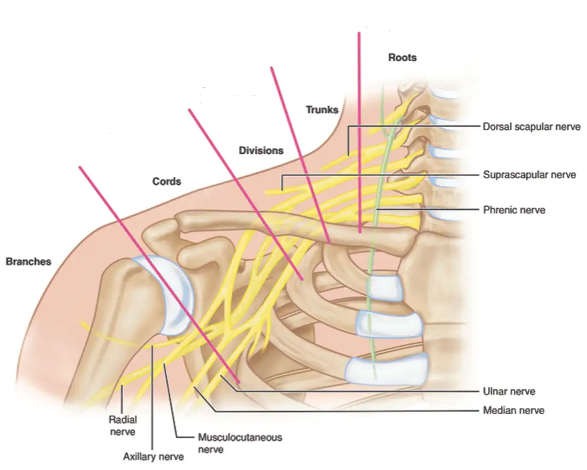
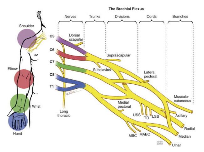
The brachial plexus is formed from five nerves that start in the spinal cord at the neck. The plexus is where these five nerves interconnect and continue on as the nerves to the muscles and skin of the arm and hand. These are the nerves that permit movement and provide feeling to the skin. There is a brachial plexus on each side of the body.
Each of the five nerves in the brachial plexus branches off into multiple other nerves that control specific functions. The location of the nerve injury within the plexus, therefore, determines which functions are lost and is important for predicting outcomes and planning treatment.
The five nerves of the brachial plexus are also divided into 5 anatomic sections. Injuries to the brachial plexus can occur in one or more of these areas:
- Spinal nerves (roots)
- Trunks
- Divisions
- Cords
- Branches
Causes of Brachial Plexus Injuries
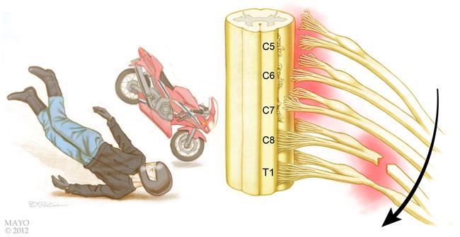
Most traumatic brachial plexus injuries occur when the arm is forcefully pulled or stretched. Many events can cause the injury, including:
- Falls
- Motor vehicle collisions
- Knife and gunshot wounds
- Most commonly, motorcycle collisions
The injury can even happen during the birthing process if the baby's shoulders get stuck coming out of the birth canal.
It is unclear how many brachial plexus injuries occur each year, but the number seems to be growing worldwide. Increased participation in high-energy sports and higher rates of survival from high-speed motor vehicle collisions may be factors in the growing number of these injuries.
Types of Brachial Plexus Injuries
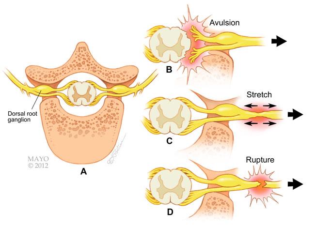
Brachial plexus injuries vary greatly in severity, depending on how many nerves in the plexus are injured and how badly.
- Avulsion. This is the most severe brachial plexus injury. The nerve root has been pulled out of the spinal cord; medically, this is known as an "avulsion" injury. These types of injuries cannot be repaired. Surgery may be used to try to improve function.
- Stretch (Neuropraxia). When the nerve is mildly stretched, it may heal on its own or require simple, nonsurgical treatment to return to normal function.
- Rupture. A more forceful stretch of the nerve may cause it to tear partially or fully. These types of injuries can sometimes be repaired with surgery.
Upper-Trunk Palsy Injury
Upper-trunk palsy occurs when the upper two or three nerves of the plexus are injured.
- Patients with upper-trunk palsies are unable to use the shoulder to raise the arm away from the body, have weakness in the arm, and may be unable to bend the arm at the elbow.
- There may be loss of sensation in the shoulder, outside of the arm, and thumb.
- A severe upper-trunk injury may paralyze the shoulder muscles (deltoid muscle and rotator cuff), as well as the muscle in the upper arm (biceps.)
Lower-Trunk Palsy Injury
Lower-trunk palsy occurs when the angle between the arm and the chest wall forcibly widens. This may damage the lower nerves and the lower trunks.
- Patients with a lower-trunk palsy will typically maintain shoulder and elbow strength but will lose hand function. Over time, this will cause the fingers to get stuck in a claw position, and the patient will not be able to perform fine motor tasks.
- Patients also typically have hand numbness in at least the ring finger and small finger.
Pan-Plexus Palsy Injury
Pan-plexus palsy may occur if the force of the injury is extreme. In pan-plexus palsy, all the nerves are damaged. This results in complete paralysis of the arm and hand, which is often referred to as "flail limb."
Gunshot Wounds
Brachial plexus injuries may be caused by a gunshot wound.
How serious the injury is depends on the bullet's caliber, velocity, and angle of entry.
- Low-velocity bullets typically damage nerves by impacting them directly.
- High-velocity bullets can injure nerves by direct impact (bruising) or, more commonly, by shockwaves that stretch the nerve. A nerve stretching and bruising injury will often recover on its own.
Gunshot wounds may also cause artery or vein injuries that require immediate or delayed repair.
Other Penetrating Wounds
A brachial plexus injury caused by a penetrating wound, such as a knife wound (laceration), may damage or cut the nerve. This type of injury to the nerve will not typically recover on its own, prompting more immediate treatment.
Additional Injuries
Because brachial plexus injuries are typically caused by high-energy, forceful events, many patients have additional injuries. These may include artery or vein injuries, fractures to the shoulder or arm, rib fractures, a collapsed lung, bleeding into the lungs or chest cavity, spine fractures, spinal cord injury, and possibly a traumatic brain injury.
Symptoms of Brachial Plexus Injuries
Symptoms depend on the type and location of the injury to the brachial plexus as well as whether the patient sustained other injuries. The most common symptoms of brachial plexus injury include:
- Weakness or numbness.
- Loss of sensation.
- Loss of movement (paralysis).
- Pain, which may result from injury to the spinal cord where the nerve rootlets are pulled out of the cord. This pain is neuropathic in nature, which means it can feel sharp, shooting, burning, and tingling. It can be very difficult to deal with and can last for a very long time.
Brachial plexus injuries that happen at the level of the spinal cord often produce greater pain than injuries farther away from the spinal cord. These injuries may also cause a burning sensation or a feeling like an electrical shock, even if the limb has no feeling or movement and nothing is touching the skin.
Diagnosing Brachial Plexus Injuries
Patients with brachial plexus injuries must be evaluated and treated within an appropriate timeframe, typically within a few months after the injury. The longer a muscle is without nerve input, the less likely it is that the muscle will function normally in the future. This is true even if the muscle eventually recovers its nerve signals.
The exact timeframe for seeking treatment depends on the type of injury and its location.
Physical Examination
If your doctor suspects a brachial plexus injury, they will perform a comprehensive examination to diagnose the injury and determine whether you have any related injuries. They will examine all nerve groups controlled by the brachial plexus to identify the specific location of the nerve injury and its severity.
The pattern by which nerves from the brachial plexus control various muscles of the arm and hand will help your doctor identify potential sites of nerve injury. Your doctor will examine all nerve groups controlled by the brachial plexus.
In addition, some patients display specific signs that help determine the location of the nerve injury:
- Narrowing of the eye pupils, drooping of the eyelid, and lack of ability for the face to sweat (Horner's syndrome) are signs that the injury is close to the spinal cord.
- A shooting nerve-like pain when the doctor taps along the affected nerves (Tinel's sign) suggests an injury farther from the spinal cord. If the location of the Tinel's sign moves down the arm toward the hand over time, it is a sign that the injury is repairing itself.
During the physical examination, your doctor will also assess your arm and shoulder for stability and range of motion.
Imaging Tests
X-rays. X-rays help evaluate bones. Your doctor will likely get X-rays of your chest, neck, shoulder, and arm to look for fractures and dislocations.
Chest X-rays help your doctor look for rib fractures or lung injury. If you are not able to take a full, deep breath during the chest X-ray, your doctor may consider pulmonary function testing with the help of a pulmonologist to rule out damage to the nerves that control deep breathing.
Magnetic resonance imaging (MRI) scan. An MRI scan is used to look at tendons, muscles, and nerves. Imaging of your neck, shoulder, and brachial plexus helps your doctor diagnose brachial plexus and any other injuries.
Computed tomographic (CT) scan. A CT scan may be used as well, often with contrast dye around the spinal cord, to look for spinal nerve avulsion (pull-out) injuries.
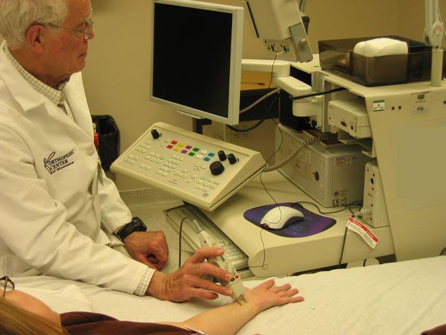
Electrodiagnostic studies. These tests measure nerve conduction and muscle signals. They are important evaluation tools because they can confirm the diagnosis, locate the nerve injury, see how serious it is, and assess the rate of nerve recovery.
A baseline electrodiagnostic examination can be done as early as 3 to 4 weeks after the injury but is more useful after several months. Electrodiagnostic studies may be repeated after the initial study to see if the nerves are recovering.
Nonsurgical Treatment of Brachial Plexus Injuries
Many injuries to the brachial plexus will recover spontaneously without surgery over a period of weeks to months, especially if they are mild. Nerve injuries that heal on their own tend to have better functional outcomes. If your doctor believes that the injury has a good potential for recovery without surgery, they may delay procedures and simply monitor your injury.
The process of the nerve healing itself takes time, and your doctor may recommend physical or occupational therapy to prevent joint and muscle stiffness and allow for healthy nerve gliding.
Surgical Treatment of Brachial Plexus Injuries
Surgical treatment is typically recommended when the nerves will not recover well enough on their own to restore necessary function to the arm and hand. It is important to note that depending on how serious the injury is, even surgery may not return the arm or hand to normal.
Things To Consider
Recovery
During your discussion with your doctor, it will be important to set realistic goals and expectations for surgical treatment. Nerves heal slowly. The recovery period after surgery is often long and requires a strong commitment to a comprehensive rehabilitation program to restore physical abilities. This is something you should consider when making the decision about whether to proceed with surgery.
Candidates for Surgery
Although brachial plexus surgery can help to restore function in many patients, there are some factors that prevent a patient from being a candidate for surgery:
- Having joint stiffness and contractures
- Advanced age
- Additional injuries or medical conditions
- Having a traumatic brain injury or spinal cord injury
- Being a smoker
Your doctor will talk to you about whether you are a candidate for surgery. Even if there is a major injury with severe loss of function, there may not be good surgical options. It is important to have realistic expectations.
Complications
- It is possible that surgical treatment will not restore desired movement or that the surgical wound may become infected. Both of these outcomes could require further surgery.
- Patients with pre-existing medical problems have additional potential risks related to any large reconstructive surgery, including chronic pain, blood clots, heart attack, stroke, and even death.
Surgical Procedures for Brachial Plexus Injuries
Several surgical techniques are used to treat nerve injury, depending on the type of injury and the length of time that has passed since the injury.
In most procedures, the surgeon makes an incision near the neck above the collarbone. If the injury extends down the brachial plexus, the surgeon may need to make another incision at the front of the shoulder. To repair or reconnect nerves, surgeons often use high-powered microscopes and small, specialized instruments.
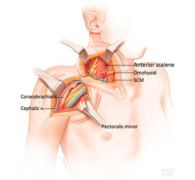
Nerve repair. In this procedure, the surgeon reattaches the two torn edges of a severed nerve. Nerve repair is typically performed immediately for sharp lacerations to the nerves, such as from a knife wound.
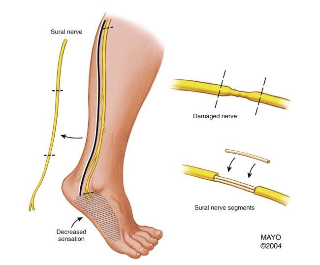
Nerve graft. Nerve grafting is a procedure in which a healthy nerve taken from another part of the body is sewn in between the two ends of a ruptured nerve. The transplanted nerve acts as a scaffold to support the injured ends as they regenerate and grow back together. Nerve grafting can be performed only if there is a functioning nerve stump at the spinal cord to conduct a nerve signal. The goal is for the transplanted nerve to guide nerve regrowth and ultimately restore nerve signals to the paralyzed muscles.
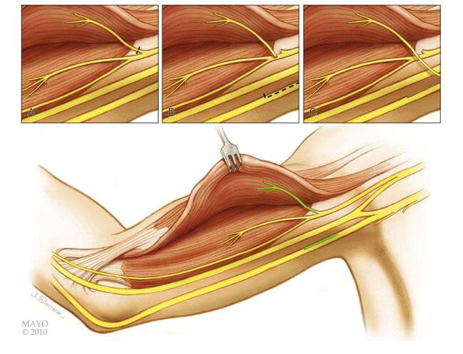
Nerve transfer. A nerve transfer procedure is used:
- When there are no functioning nerve stumps in the neck to which nerve grafts can be connected, or
- The surgeon thinks it will result in better recovery than grafting
In this procedure, a healthy donor nerve is cut and reconnected to the injured nerve to provide a signal to a paralyzed muscle. In many cases, the healthy nerve is connected closer to the affected muscle. In other cases, the healthy nerve is connected to the damaged nerve within the brachial plexus.
Tendon and muscle transfers. Surgery that focuses on rebuilding the tendon (tendon transfer) or muscle (free-functioning muscle transfer) may be a better option for:
- Patients who do not recover well enough, with or without nerve surgery
- Patients who seek care more than 12 months after the injury
A tendon transfer is procedure in which the tendon of a functioning muscle is cut and sewn into a nonfunctioning muscle tendon to restore a specific motion or motor function.
In a free-functioning muscle transfer, a muscle from one part of the body is moved to the injured area, along with its tendon, artery, vein, and nerve. Each of these structures is connected to the matching structures in the injured area to restore motion or motor function.
Recovery and Rehabilitation After a Brachial Plexus Injury
Because nerve regeneration occurs slowly at a rate of approximately 1 mm/day, recovery from a brachial plexus injury takes time. In fact, patients may not experience results for several months. A positive mindset and the support of family, friends, and healthcare professionals are important to recovery and rehabilitation.
During this recovery process:
- Occupational therapists teach patients how to use the unaffected arm to perform daily activities like eating and personal hygiene.
- Physical therapy of the shoulder, elbow, wrist, and fingers involves specific exercises to prevent stiffness, contractures, or muscle atrophy.
- A physical therapist can also recommend assistive devices, such as splints or braces, to help support a limp arm and joints.
- Compression gloves and sleeves may be used to prevent swelling in the affected arm, which can lead to pain and joint contractures.
- Pain may be managed with medications, therapy, and/or assistive devices.
- Patients will require healthy coping skills to adjust their lives — including work and daily activities — around a less functional arm and/or hand.
Future Developments
Although brachial plexus injuries can be devastating and hard to manage, a team approach to treatment has led to significant improvements in patient function. Current technology cannot return patients with a flail limb to their pre-injury state, but restoring some function to the arm is a major advance.
Future developments in treatment of these injuries include newer techniques for repairing or transferring nerves, as well as new medications or materials to help promote and stimulate healthy nerve regeneration.

Contributed and/or Updated by
Peer-Reviewed by
AAOS does not endorse any treatments, procedures, products, or physicians referenced herein. This information is provided as an educational service and is not intended to serve as medical advice. Anyone seeking specific orthopaedic advice or assistance should consult his or her orthopaedic surgeon, or locate one in your area through the AAOS Find an Orthopaedist program on this website.
The content on this website may have been translated from English into other languages using artificial intelligence tools. The translations are provided for the convenience of readers and may not been reviewed or verified by medical professionals. AAOS makes no guarantees regarding the accuracy, reliability, or completeness of the translated information and disclaims all liability for any issues arising from its use. Users are strongly encouraged to refer to the original English content and consult qualified healthcare professionals before making any medical decisions. By using the translated content, you acknowledge and accept these limitations and assume full responsibility for any reliance on the information provided.